Search "Comfort Keto" Website
Search this site
512 results found with an empty search
- How Do You Like Your Juicy Sirloin Steaks Hot Off A 3D Printer?
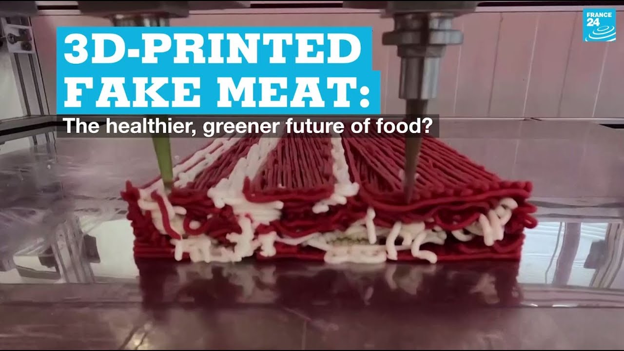
Things are getting weirder every passing day. Faux meat will soon be on the menu at steak houses and other restaurants this year. After securing $29 million funding, an Israeli start-up, Redefine Meat Ltd. is now ready to target steak houses and other restaurants worldwide with its 3D-printed facsimiles of beef cuts, from fillet to rump and brisket. The company wants to change the "belief" that delicious meat can only come from animals. We wonder, where else can meat come from? What kind of craziness is it, that whatever is printed on a 3D printer can be called "meat"? Food giants like Nestle and McDonald’s Corp. along with multi billion dollars venture-capital investors call this charlatanry (charlatan is a person or a company practicing quackery in order to obtain money, fame, or other advantages through deception) "alternative-protein" as the so-called environmental and health (???) concerns supposedly entice consumers to fake products like faux burgers or nuggets. While investment is flowing into fake meats, the producers of this quackery admit that more needs to be done to improve the taste of products and lower prices to compete with conventional meat. Redefine is trying to take plant-based products to the next level by giving diners the same sensory experience as eating prime beef. Meanwhile, other opportunist charlatans are popping up elsewhere. Israeli startup, Aleph Farms Ltd., unveiled the world’s first slaughter-free ribeye steak. Barcelona-based Novameat Tech plans to start selling 3D-printed vegan meat to restaurants in Europe this year as it raises funds for a production scale-up. 3D printers loaded with plant-based “ink” can print the meat countless times and deliver a complex layering of muscle and fat to recreate the right texture. We bet the ink cartridges are not that cheap.
- Reprogram Your Mind To DESTROY LAZINESS & Improve Focus TODAY!
At one point or another, like any one else, you felt stuck in a rut with feelings of laziness and fear. During these times it can be really hard to take a step back and wonder what’s happening in the body, as opposed to just the mind, but it’s the link between the two that can push us through it. Dopamine Is the Key Neurotransmitters have incredible power over how the body and mind function. Let's look at the chemical symphony happening in the body all the time. Dopamine is responsible for craving, motivation, and pursuit. Adrenaline relates to agitation and endurance. Serotonin helps us be grateful and feel good about what we have. And, acetylcholine can helps focus. You can react to the world around you and make positive changes by purposefully leveraging these inner reactions. In this Podcast, Dr. Andrew Huberman talks about opening the window into the body's own inner neurochemistry, training the nervous system, and confronting the body's inner real estate. Dr. Andrew Huberman is a neuroscientist and tenured Professor in the Department of Neurobiology at the Stanford University School of Medicine. He has made numerous important contributions to the fields of brain development, brain function, and neural plasticity, which is the ability of our nervous system to rewire and learn new behaviors, skills, and cognitive functioning. He is a McKnight Foundation and Pew Foundation Fellow and was awarded the Cogan Award in 2017, which is given to the scientist making the largest discoveries in the study of vision. His lab’s most recent work focuses on the influence of vision and respiration on human performance and brain states such as fear and courage. Work from the Huberman Laboratory at Stanford University School of Medicine has been published in top journals including Nature, Science, and Cell and has been featured in TIME, BBC, Scientific American, Discover, and other top media outlets. The Podcat dives into: - The connection between fear, laziness, and motivation (0:40) - The difference between dopamine, adrenaline, and serotonin (13:35) - How to increase dopamine (15:04) - How to get your mind to stop racing (26:41) - An exercise to try if you are having a hard time falling asleep (31:32) - The benefits of hypnosis (34:41) - What to do if you are having trouble focusing (48:32) - Why structure is one of the best ways to create freedom (57:17) - How our phones are eroding our creativity, relationships, and more (1:01:37) - The importance of having times of no focus each day (1:12:24) For more on Dr. Andrew Huberman, you can follow him on Instagram @hubermanlab, and through his website http://www.hubermanlab.com/.
- Start Your Weight Loss Journey NOW With These Two Simple Steps
First Things First: Drink Enough Water! Did you know that about 75% of the human body is composed of water? And did you know that if you ask most people what they would like to drink water would be on a last choice? Because your body is composed of about 75% of water, that means that it needs water to function optimally. This is one of the easiest tips to implement yet most people neglect it. If you just start drinking more water every day it will go a long way in speeding up you fat loss. Researchers in Germany have found that people who drank water had an increase in the amount of calories they burned per day. How Much Water Should You Drink? We suggest that you start with two glasses a day and then increase at your own pace until you reach ten glasses per day. We recommend that you follow this very simple water consumption formula: Determine your body weight in pounds, divide it by two and drink that many in ounces of water each day. For example, a 200 lbs person should drink 100 ounces of water each day. That is almost 2/3 of a gallon per day... Recommended product: This BPA free, reusable 100 oz water bottle with straw has "Daily Water Intake Tracker" marked on it. This bottle's time marker feature from 7 am to 7 pm will help you track your daily water intake. At the same time, it acts as a reminder, and works well helping you to adopt a healthy hydration habit. With this water bottle, you will surely meet your goal for drinking enough water throughout the day. Avoid Spiking Of Your Insulin Level You can eat lettuce and work-out all day long and never lose any weight if you don't balance your blood sugar and hormones, in particular INSULIN. But that's extremely challenging for the majority of people (as long as they are not in ketosis), because most folks living in the modern world are desperately addicted to sugar. The addiction to sugar is real and many experts tell that it is more difficult to quit sugar than smoking, alcoholism, or even addiction to "hard drugs." Some experts go even so far claiming that sugar is much more addicting than heroin. If you feel like you can't give up sweets, or you can't give up dessert, or you can't give up pizza, pasta or bread (these are broken down nearly instantly into sugar)... then you know exactly what I'm talking about. And if you think that's bad... it's actually even worse: Because it's now been proven that synthetic sweeteners that many people rely on to help them reduce sugar in their diets are actually worse for you. Studies after studies have shown that these zero-calorie synthetic sweeteners actually trigger chemical reactions in the body that cause you to produce more fat and gain more weight. But if you can get off the sugar roller-coaster, you will see instantaneous changes in your weight and in your physic. Here are 3 things that can help you lose weight now... When you consume foods that have sugar, high fructose corn syrup, maltose, or any of the other names used to disguise the word "sugar" on ingredient labels... or... when you eat white or refined starches like bread, pasta, rice, or potatoes... you cause your insulin levels to spike. The insulin spikes leave you feeling fatigued, depressed, and cause your body to store fat. They also cause your blood-sugar level to crash... which is why you immediately get cravings to eat more. A good number of people spend their whole day trapped in this cycle. Natural Sweetener With Zero Calories: Stevia The natural sweetener stevia leaf has no calories at all... yet is extremely sweet. Stevia is also quite a bit sweeter than sugar. Numerous studies on the safety of stevia show that, as a natural product, it is extremely safe and non-toxic. And it has been used in many cultures from Paraguay to Japan as a health-promoting herb. This miracle herb can used to sweeten anything from your morning coffee to candy as a healthful substitute for sugar.
- HEALTHY LIVING Tips to LIVE LONGER and Feel Better
You may think of insulin resistance as being reserved for diabetics, and you may have even drawn to the conclusion, that the problem is glucose being too high. In this video, Dr. Bikman, a scientist that studies metabolic disorders caused by insulin resistance, explains in very simple terms that insulin resistance can still develop even in people with seemingly normal glucose levels. Dr. Bikman’s explanation of the complicated nature of insulin in the body and his unique take on facts that can literally save your life with knowledge are exactly what we need more of. Whether you consider yourself to be healthy, prediabetic, or struggling with other health issues this episode may give you the insight you need today.
- Burn Fat & Reverse Insulin Resistance | Dr. Benjamin Bikman Explains Metabolic Flexibility
Want to lose body fat, improve energy levels, and better understand how your metabolism works? In this informative lecture, metabolic scientist Dr. Benjamin Bikman (Ph.D. in Bioenergetics & Metabolism) explains how the body chooses between burning sugar and fat for fuel, and why insulin plays a central role in metabolic health, weight management, and overall well-being. Although this presentation was recorded several years ago, the science remains highly relevant today. Dr. Bikman breaks down complex metabolic concepts into easy-to-understand principles that can help you make more informed decisions about nutrition, fasting, and long-term health. In this presentation, you'll learn: ✅ How insulin influences fat storage and fat burning ✅ What insulin resistance is and why it matters ✅ The difference between metabolic flexibility and metabolic inflexibility ✅ How the body transitions into a fat-burning state ✅ Why ketones are an efficient fuel source for the brain, heart, and muscles ✅ The role of fasting and nutrition in supporting metabolic health 🔍 Topics Covered: • Metabolism and energy production • The three macronutrients: carbohydrates, protein, and fat • Sugar versus fat as fuel • Understanding insulin and its functions • Insulin resistance and metabolic dysfunction • Metabolic flexibility and fuel switching • Entering the fat-burning zone • Ketones and their potential benefits • Intermittent fasting concepts • Foundational nutrition principles for metabolic health About Dr. Bikman Dr. Benjamin Bikman is an internationally recognized researcher specializing in insulin resistance, metabolic disorders, and fat metabolism. He is the author of the bestselling book Why We Get Sick and has become one of the leading voices in the field of metabolic health and nutrition science. This episode dives into: 📌 Chapters 0:00 - Intro 1:03 - What is Metabolism? 1:32 - The 3 Macronutrients 2:01 - What Fuel Does Our Body Use? 2:49 - What is Insulin? 3:08 - How Food Affects Insulin 5:06 - How to Burn Fat for Fuel 6:33 - What is Insulin Resistance? 8:14 - What Happens When We Eat Carbs? 8:48 - Metabolic Flexibility Explained 10:42 - Metabolic Inflexibility & Why It Matters 11:37 - Fuel Tanker Analogy 17:38 - Unicity Nutrition Principles 18:22 - What is 4-4-12 Fasting? 18:43 - Entering the Fat Burning Zone 20:04 - What Are Ketones? 21:03 - Benefits of Ketones 22:58 - The Three Nutritional Principles 28:58 - Conclusion You can follow Dr. Bikman on Instagram @BenBikmanPhD, on Facebook @BenjaminBikmanPhD, on Twitter @BenBikmanPhD, and through his websites https://cell.byu.edu/bikman-lab/ and https://www.insuliniq.com/. You can find his book, Why We Get Sick: The Hidden Epidemic at the Root of Most Chronic Disease―and How to Fight It, right here: https://benbellabooks.com/shop/why-we-get-sick/
- Insulin, Brown Fat & Ketones With Benjamin Bikman, PhD
In this really good video, Dr. Ben Bikman shares his latest scientific findings between the ketogenic diet and brown adipose tissue activation. Connect with Dr. Ben Bikman ➢ https://bikmanlab.byu.edu ➢ http://www.insuliniq.com Key Takeaways 02:02 During PhD studies, he learned that insulin is the metabolic key. Now that he has his own lab, the relevance of insulin upon normal metabolic health is his area of study. 03:11 Insulin is needed to Grow Fat Cells. 03:35 Distortion of the Fat Cell 05:59 As we grow larger fat cells and we reach 150% of what is ideal for our bodies, we start making new fat cells. As someone loses weight, they will not reduce the number of fat cells. They shrink the size of their existing cells. 07:14 Triglycerides are Not Relevant to Insulin Resistance 09:34 Muscle is Our Biggest Glucose Sink 10:35 Insulin is a Factor in Inflammation 12:08 Exercise is Important, but Diet is Critical 13:45 Toxic Lipids Cause Mitochondrial Alterations 15:39 Brown Adipose Tissue and an Uncoupling Protein 18:39 When Brown Fat Cells are Exposed to Insulin, Their Metabolic Rate Will go Down by about Half. 19:38 When our White Fat is Exposed to Ketones, Our Fat Goes from Storing to Wasting. 20:34 The Evolutionary Benefit of Ketones Inducing Wasting: Perhaps the conversion of white fat into heat burning instigated by ketones was meant for heat production. Our bodies act protectively. Evolutionarily, ketogenic diets would be seasonal. 24:55 Cold Thermogenesis to Activate Brown Fat: Constant cold exposure, to the point where you are shivering, works to activate brown fat. Ice baths and genuine cold exposure does this as well. 27:31 You Can Alter Your Metabolic Rate 30:24 Benefits of Low Insulin 32:46 We Need Insulin: It is necessary for normal mitochondrial function. Eating a healthy low carb diet will give you enough insulin variability. We want to keep insulin normal. 25:16 Time Restricted Feeding and Fasting to Trigger Beiging 28:05 We are All Insulin Resistant First Thing in the Morning 39:01 The Effect of Protein and Carbs on Insulin: If you are insulin resistant, you will get a huge long lasting fat storage spike from eating something like a bagel, as opposed to the quick bump for someone who is insulin sensitive. 41:45 Speculation that Exogenous Ketones Turn to Fat when Insulin is high 46:34 Dr. Bikman’s Morning Routine 48:08 Dr. Bikman’s Favorite Exercise is the Deadlift 49:09 Dr. Bikman’s Desert Island Herb, Nutrient or Botanical: He would bring cow liver, which is packed with mitochondria. We do not adopt the mitochondria of the meat we eat, but we can get the building block components of the mitochondria. 51:06 Dr. Bikman’s Elevator Pitch: Stop emphasizing a high carbohydrate/low fat diet. 51:36 Don’t Worry about Gluconeogenesis
- Antioxidant Rich Foods
The vitamin C and vitamin E, are known to protect the body against the destructive effects of free radicals. Antioxidants neutralize free radicals by donating one of their own electrons, ending the electron-"stealing" reaction. The antioxidant nutrients themselves don't become free radicals by donating an electron because they are stable in either form. They act as scavengers, helping to prevent cell and tissue damage that could lead to cellular damage and disease. Vitamin E Vitamin E is the most abundant fat-soluble antioxidant in the body. Vitamin E is one of the most efficient chain-breaking antioxidants available. It is the primary defender against oxidation and lipid peroxidation (creation of unstable molecules containing more oxygen than is usual). Vitamin C Vitamin C is the most abundant water-soluble antioxidant in the body. Vitamin C acts primarily in cellular fluid. It is particularly beneficial in combating free-radical formation caused by pollution and cigarette smoke. It also helps return vitamin E to its active form.
- Healing from the Garden: Natural Herbs for Pain Relief, Immunity & Everyday Wellness
Many people can reduce their reliance on pills and medications by learning the basics of natural plant remedies, which may help support pain relief without many of the unwanted side effects associated with conventional medications. In this short blog post, we’ll introduce several common herbs and plants that can be easily incorporated into your daily diet to help manage minor aches and discomforts. Best of all, many of these beneficial herbs can be successfully grown right at home. Herbs and plants can be easily incorporated into your daily diet to help manage minor aches and discomforts. Aloe Vera Place aloe vera in a spot with plenty of sunlight and water regularly. Aloe vera is widely known for its soothing and healing properties, especially when it comes to minor scrapes, bruises, and sunburns. Its cooling gel helps reduce inflammation and supports the skin’s natural healing process. When it comes to growing and caring for aloe vera, place it in a spot with plenty of sunlight and water it regularly—but be careful not to overwater, as it prefers well-drained soil and can be sensitive to standing moisture. Basil (Ocimum basilicum) Basil is a natural remedy for headaches. Basil isn’t just a flavorful addition to meals—it’s also a natural remedy for headaches. To harness its soothing effects, try rubbing freshly crushed basil leaves on your temples. Alternatively, steep the leaves in boiling water and use the infusion as a calming foot soak. Basil grows best in pots and prefers plenty of sunlight. To keep the plant from becoming bitter, regularly pinch off the white flowers as they begin to form. Lavender (Lavandula) Lavender is well known for its anti-inflammatory properties. Lavender is well known for its calming scent, but it also boasts notable anti-inflammatory and skin-soothing properties. Beyond aromatherapy, lavender has traditionally been used as a natural topical remedy for minor skin irritations and blemishes. When properly diluted, lavender oil may help reduce redness, calm inflammation, and support clearer-looking skin when applied directly to affected areas. Lavender is not commonly consumed as a food herb like parsley or basil, though certain culinary varieties are occasionally used in teas, desserts, and herbal blends in small amounts. Historically, lavender has been more widely valued for its fragrance, medicinal uses, and natural insect-repelling properties. Lavender helps protect clothing made from natural fibers because its strong natural oils repel moths and other fabric-damaging insects. For centuries, dried lavender bundles and sachets have been placed in closets, drawers, linen chests, and wardrobes to help repel moths and other fabric-damaging insects that target natural fibers such as wool, linen, and cotton. This traditional practice remains popular today among people seeking natural alternatives for protecting valuable clothing and household textiles. Lavender thrives in light, well-draining soil and requires plenty of sunlight. Proper drainage is essential for keeping this fragrant herb healthy and productive. Lemon Balm (Melissa officinalis) It’s best to plant lemon balm in a pot. It can easily take over your garden if left unchecked. Tired of cold sores making unwanted appearances? Lemon balm is a natural remedy that helps prevent and heal these pesky blemishes. Its antiviral properties make it especially effective. You can also rub the leaves directly on your skin as a gentle, natural insect repellent. When growing lemon balm, it’s best to keep it in a pot—this hardy herb spreads quickly and can easily take over your garden if left unchecked. Mint (Mentha Piperita) It’s best to plant mint in a pot. It can easily take over your garden if left unchecked. Mint is a versatile herb with a wide range of natural healing benefits. A tea made from fresh mint leaves can help relieve anxiety, nervousness, flatulence, and stomach cramps. Mint is also excellent for steam inhalation—simply place a handful of leaves in a bowl of boiling water, drape a towel over your head, and breathe in the steam to help clear sinus congestion. When growing mint, it’s best to use a sunken pot or container, as the plant spreads rapidly and can easily overrun garden spaces if left unmanaged. Parsley (Petroselinum crispum) If you find yourself frequently getting sick, try eating a teaspoon of fresh parsley daily. Parsley is more than just a garnish—it’s a powerful immune system booster. If you find yourself frequently getting sick, try eating a teaspoon of fresh parsley daily. It’s also a natural breath freshener; simply chew a few leaves to help combat bad breath. Parsley thrives in moist, well-watered soil. Be sure not to let the soil dry out, as this plant prefers consistently damp conditions to grow successfully. Rosemary (Rosmarinus officinalis) A unique winter remedy: infuse rosemary with red wine, cloves, and cinnamon to combat the effects of winter colds. Rosemary is a fragrant herb known for its mood-lifting and healing properties. It has been traditionally used to help reduce symptoms of seasonal affective disorder (SAD) and even ease hangovers. One of the most common ways to enjoy its benefits is by brewing a tea with fresh rosemary leaves. Here’s a unique winter remedy to try: infuse rosemary leaves with red wine, cloves, and cinnamon to create a warming tonic that may help combat the effects of winter colds. Rosemary thrives in full sunlight and does best when given plenty of space and well-drained soil. Sage (Salvia officinalis) Sage is a natural remedy for throat discomfort. Have a sore throat? Sage has long been used as a natural remedy for throat discomfort. Simply steep fresh sage leaves in hot water to make a soothing herbal broth. Once cooled, gargling with this infusion can help ease inflammation and reduce soreness. Sage prefers full sun and grows best in dry, sandy soil. It’s a hardy plant that thrives with minimal watering once established. Thyme (Thymus vulgaris) Thyme is a potent antioxidant and antiseptic. Thyme is a potent antioxidant and antiseptic with a range of medicinal benefits. Drinking thyme tea before bed can help alleviate cold symptoms and promote a restful night’s sleep. However, it’s important to note that thyme should be avoided during pregnancy. Thyme thrives in dry, light soil and requires plenty of sunlight to grow successfully.
- Cortisol: Reason For The Squeaky Wheels In Your Life
When you have a squeaky wheel, how do you solve the problem? “The worst wheel of the cart makes the most noise.” ~ Benjamin Franklin Do you complain about it? Do you grease it up? Do you take it apart to find out why it started squeaking in the first place? Or simply ignore it and keep pushing the cart along? What’s worth more to you — a temporary patch or a permanent fix? Sure, putting grease on the wheel might temporarily stop it from squeaking. But it doesn’t solve anything long-term. How many squeaky wheels do you have in your life right now? And lastly, how long have your squeaky wheels been squeaking? Although may you experience naturally high levels of cortisol from time to time throughout your life, long term excessive levels of cortisol hormone floating throughout your body may cause serious health problems. Every squeak is a symptom which needs to be addressed, if you want to lead a reasonably healthy life. Cushing’s syndrom is caused by long term excessive levels of cortisol hormone floating throughout your body due abnormal function of adrenal and pituitary glands. This condition may also cause adrenal fatigue. Although many people experience at least somewhat high levels of cortisol at one time or another, true Cushing’s disease is rare compared to other endocrine disorders. This can make it hard for patients to get an accurate diagnosis. Especially woman of ages between 25–40 years (about 70 percent of all Cushing's patients) may be dealing with myriad symptoms that just won’t go away no matter what they try or what their doctor prescribes. There’s a high probability that such folks may have Cushing’s disease. Even if you aren’t in that demographic, you can’t rule this condition out, as it can affect anyone. People suffering from Cushing’s disease are often misdiagnosed without identifying what’s causing their symptoms. They might be brushed off by simply being told to “diet and lose weight.” Yet, misunderstandings about Cushing’s disease can further increase anxiety, self-blame and chronic stress, which only worsens the underlying issue. Most common Cushing’s syndrome symptoms: persistent weight gain (especially in the abdominal area, lower back, upper body or near the neck, which is sometimes called a “buffalo hump“) a puffy, rounded face skin problems, including redness, dryness and acne slow healing stretch marks fatigue, muscle weakness high blood pressure high blood glucose (sugar) levels changes in mood, including depression and anxiety irregular periods, fertility problem, slow sex drive abnormal hair growth on the body and face (especially noticeable in women) weakened bones, higher risk for fractures or osteoporosis digestive problems, constipation bloating/water retention kidney stones trouble sleeping The good news is through diet and lifestyle changes, you can naturally treat this adrenal condition. Foods to add to your diet in order to overcome adrenal fatigue (because they’re nutrient-dense, low in sugar and have healthy fat and fiber): Coconut Olives Avocado and other healthy fats Cruciferous vegetables (cauliflower, broccoli, Brussels sprouts, etc.) Fatty fish (e.g., wild-caught salmon) Free-range chicken and turkey Bone broth Nuts, such as walnuts and almonds Seeds, such as pumpkin, chia and flax Kelp and seaweed Himalayan sea salt Fermented foods rich in probiotics Chaga and cordyceps medicinal mushrooms Other Natural Ways of Lowering Cortisol Levels Essential oils such as lavender, myrrh, frankincense and bergamot are also helpful for fighting stress. These are capable of lowering cortisol, reducing inflammation, improving immunity, balancing hormones, and helping with sleep and digestion. Natural adaptogen herbs help tremendously with lowering cortisol levels. They boost your ability to deal with stress. They also have energizing qualities, antioxidant effects, antidepressant effects, and can help naturally lower fatigue, blood pressure and blood sugar levels. If you’re dealing with Cushing’s disease, it’s best to talk to your doctor before using any herbal treatments, especially if you’re taking medications, but in general these plants have been used safely for thousands of years with little side effects. There are many different proven adaptogenic herbs that can help lower cortisol, including: ashwaganda astragalus ginseng licorice root holy basil medicinal mushrooms, including reishi and cordyceps rhodiola Read more about Cushing's Syndrome here.
- How to Stock Your Fridge If You Want Your Food to Last!
One of the most fundamental ground rules for keeping refrigerated items fresh: “The colder the fridge, the longer things will last.” Refrigerators should be set between 35 and 40 degrees Fahrenheit for both safety and quality—even a degree can make a difference. Due to the frequent opening and shutting of the refrigerator door, the food in the front and on the side of the refrigerator door may be most likely well above 40 degrees. So, make sure your fridge is set cool enough and push things as far back from the door as possible. Also, follow this link to find out which foods you possibly didn’t know that you could freeze. And here, you will find out here how long different types of meats last in the fridge. We rounded up a few tips which will help you make sure your food lasts longer and save you from having to make extra grocery store trips. Choose foods with staying power! Some long-lasting picks: eggs (they’re good up to five weeks, and it’s easy to know when one’s gone bad), winter squash, citrus, onions, hard cheeses (avoid low-sodium cheese which won’t last as long), pickles, cream cheese, sour cream, heavy cream, and bacon. Separate your produce! All fruits and vegetables are not created equal, and tossing them all into the same crisper drawer can significantly shorten the lifespan of some food items. Tomatoes for example, produce a lot of ethylene gas, a plant hormone that speeds up the ripening process. Asparagus, garlic, onion, broccoli, cauliflower, carrots, green beans, peppers, and strawberries are among the produce items most sensitive to the effects of ethylene. If you must refrigerate these items, store high ethylene producer tomatoes in its own drawer. These foods should be stored at room temperature! Tomatoes, onions, and avocados last longer at room temp. Here are some foods that shouldn’t go in your fridge. Find out why: Basil, cucumbers, onions, tomatoes, coffee, garlic, hot sauce, honey, sliced low carb bread, nuts, avocados, olive oil, bell peppers, pickles, chocolate, eggplant. Don't wash everything right away! Moisture leads to mold, so washing a box of strawberries at once, for example leads to rapid spoilage. Instead, only wash the amount you’re going to eat in one sitting immediately before eating it and leave the rest dry. Maintain proper moisture levels with your fresh herbs for them to last longer Fresh herbs make a big impact in all kinds of recipes, yet those delicate greens never last long. Growing your own is always a good option, as is using dried. The best way to extend the life of cut leafy herbs like cilantro and parsley is to maintain proper moisture levels by sticking them in a jar of water and placing a plastic bag loosely over the top. Of course you should change the water regularly. Here’s more information on how to store your herbs properly. Minimize your cut surfaces Exposure to oxygen is one of the fastest ways to spoil food. When you need half only an onion, tomato, or lime, you want to minimize your cut surfaces (i.e. don’t chop the rest of that onion) and cover it tightly with plastic wrap. This will also prevent odors from stinkier items like onions from permeating other foods in the fridge. Don’t wrap your cheese in a plastic foil Cheese is a living food. The common yet WRONG way of storing a cut wedge of cheese is to wrap it in a plastic foil. This traps moisture inside and doesn’t let the cheese breathe. That's why cheese starts to feel slimy before sprouting bad kind of mold. Cheese should be wrapped in parchment or wax paper and placed in a small Tupperware container with a tight-fitting lid. Wrap right Wrap your leftovers in plastic wrap before popping then in a Tupperware container. This way, you will prevent freezer burn in case you plan to keep them for longer periods. When you don’t want to take any chances with food going bad before it’s time, sucking the out of the plastic bag your leftovers are in is the best solution. Vacuum sealing can keep food fresh for five times as long as other storage methods. Enjoy your perfectly fresh kept left-overs. Bon appétit! Let's Celebrate Spring in a Ketogenic Way!!! During the peak of the pandemic, we served our repeat customers only! Now that Spring is here and the pandemic is under control, we decided to reboot! We are now accepting orders from "6" NEW CUSTOMERS from SAN JACINTO and HEMET area. The orders will be accepted on "first come, first served" basis.Please place your order for our chef prepared ketogenic menu of the week on our website at https://www.myketopal.com/comfort-keto-meals by Friday midnight in order to receive your weekly prepared keto meal package on following Monday! FREE curb-side pickup in San Jacinto; Delivery within 30 mi. radius from San Jacinto available for $10.
- Here Is What You Do If You Have High Blood Pressure?
High Blood Pressure is typically caused by stress, nutrient deficiencies and lack of hydration at a deeper level. High Blood Pressure can be combated by leading a ketogenic lifestyle, introducing healthy fats into your diet, and systematically reducing your stress levels. If not treated with the right kind of nutrition (low carb high fat, LCHF), High Blood Pressure may lead to blood thickening, thus cause the heart to work harder in order to get the blood flow at acceptable levels. Also, strokes are typically caused by blood thickening. Hydration Water hydrates at a surface level, add a few drops of organic lemon and it will penetrate the cells at a deeper level and provide more hydration for the cells. Nutrition Daily regime must consist of meals that do NOT contain industrially produced fats (margarine, canola oil, vegetable oil), refined sugars, and any chemicals (dyes, flavoring, preservatives, etc.). We suggest that you consume as much as possible pesticide-free non-GMO local grown vegetables. Have MORE antioxidants, vitamins, minerals which help with brain function. Below is a few more dietary suggestions to combat issues resulting from High Blood Pressure. NATURAL BLOOD THINNERS Turmeric (curcumin, the active ingredient, is anti-inflammatory and blood thinning) Ginger (also anti-inflammatory, natural occurring acid salicylate which is a potent blood thinner) Vitamin E (400-800 IU per day, also found in almonds and sunflower oil, will aid in SKIN health) Garlic (natural anti-biotic and antithrombotic that reduces blood clot formation, also boosts the immune system.) Cinnamon (toxic if not organic as it [just like stevia] absorbs toxins and pesticides which is why it is such an amazing plant as it absorbs toxins in the body and flushes them out, thus best had organic. It contains coumarin, powerful for blood-thinning.) Grape Seed Extract (Contains antioxidants to protect blood vessels and prevent high blood pressure as well as aiding overall blood health) LOWER CHOLESTEROL Avoid trans fats or saturated fats. Instead: Monounsaturated Fats and Polyunsaturated Fats (Olive oil, Olives, Tree nuts, Avocados, Omega 3s) Eat Soluble Fiber Fish Oil Avoid refined sugars and grains Stress The number one cause of high blood pressure is stress. Stress creates nutrition deficiency just by existing. Whatever stresses you have from the past memories or so forth may be a part of the health issues. Meditation Some meditation techniques can help your blood pressure decrease. It may take only 5 minutes every few hours, quick and just to calm the mind and aid any stresses you might be having. Exercise Yoga is great for full body circulation. Stretching your muscles is very helpful too. Sleep Get 8 hours of sleep nightly. Sleep early, wake early! Entertainment Whatever entertainment you are watching or reading, encourage more peaceful and happy things. Even though it might seem fun to watch scary or intense things, it can make the mind subconsciously stressed which affects the nervous system negatively. Aromatherapy Use Essential Oils that are very safe and very good for healing many issues. Smelling Orange Essence helps you during very difficult times. Acupuncture This is very helpful with overall health and can calm down the nervous system as well) Magnesium Magnesium is key for relaxation and muscle tension relief. It will also help with bowel movements. Typical Medications Given for Strokes and Their Side Effects: The below listed drugs lower the immune system, damage the skin, have allergic properties. Please note that best thing to do to heal the damage resulting from High Blood Pressure is to counter with healing methods and foods to aid the body. Other side effects of the below listed medication also possible but not listed. Clopidogrel: Blood Thinner This medication takes a great toll on the immune system and damages the immune system functions. This means the body will be more prone to infection and illness because the immune system is compromised. If you receive a rash, one of the side effects of the medication is rashes as well. The list of side effects is extremely long, I have listed some below. You will have to counter these side effects deep damaging affects with immune system boosting natural foods and a healthy diet. Declared Side Effects: Upper respiratory tract infection, chest pain, headache, flulike symptoms, joint pain, pain, dizziness, diarrhea, rash, urinary tract infection, nosebleeds, increased bleeding, itching, bruising. Other side effects include: Eczema, hepatitis, acute liver failure, low blood pressure, severely lowered white blood cell count (white blood cells are the defense cells that destroy free radicals and mutated cells). Additional side effects include: lymphatic system disorders (lymph is the key for the immune system), anemia, eye disorders, gastrointestinal disorders, general disorders, immune system disorders, nervous system disorders, psychiatric disorders, respiratory disorders, skin tissue disorders. Other side effects are also possible but not listed. Atorvastatin: Cholesterol This drug is not good for muscles or the digestive system, which means you will have to counter with pro-biotics and other natural healthy foods to repair damage. Declared Side Effects: Gastrointestinal symptoms, joint pain, loss of appetite, insomnia, indigestion, muscle aches/issues, muscle inflammation, muscle ache, lightheadedness, fainting, breathing issues. Other side effects are also possible but not listed. Lisinopril: High Blood Pressure This drug has a side effect of a rash, so you will have to take immune system boosting natural foods and healthy options which are listed above to counter the effects of this and the other drugs. This drug also has skin issues associated with its use. Declared Side Effects: Dizziness, cough, headache, high potassium levels (this can change the heart rhythm and too much of potassium can be dangerous), diarrhea, low blood pressure, chest pain, fatigue, rash, psoriasis, skin irritation, stomach pains.
- Pistachios: A Nutrient-Dense Snack with Surprising Benefits
When it comes to nuts, pistachios are often overlooked, even though they are nutritional powerhouses. These small green gems are rich in fiber, heart-healthy fats, and satiating protein. They also provide a variety of antioxidants that help combat oxidative stress and support overall health. Pistachio trees have been cultivated throughout the Middle East for thousands of years and have long been considered a prized delicacy. One unique characteristic of pistachios is that their shells naturally split open as the nut matures, making them easier to enjoy than many other tree nuts. They are used in countless recipes around the world and remain one of the most minimally processed snack foods available. Pistachios are even mentioned in the Old Testament (Genesis 43:11). How to Choose Packaged Pistachios Today's grocery stores offer a wide variety of pistachios, including raw, roasted, shelled, and flavored options. We occasionally use flavored pistachios in salads, particularly varieties seasoned with chili lime, cumin, or black pepper. When selecting flavored pistachios, we recommend checking the sodium content and choosing products that contain less than 5% of the Daily Value per serving whenever possible. If you're following a low-carb or ketogenic lifestyle, avoid honey-roasted pistachios and all other varieties sweetened with added sugars. Health Benefits of Pistachios Pistachios May Support Better Sleep Melatonin is a natural hormone that helps regulate the body's sleep-wake cycle. Pistachios contain naturally occurring melatonin, making them one of the richest dietary sources identified to date. Research suggests that pistachios contain exceptionally high levels of melatonin compared to many other foods. Consuming pistachios as part of a balanced diet may help support healthy sleep patterns. Potential benefits of melatonin include: Supporting the body's natural sleep cycle Helping the body relax before bedtime Promoting deeper, more restorative sleep Reducing the effects of jet lag Shortening the time it takes to fall asleep Providing antioxidant support Pistachios May Help Support Healthy Blood Sugar Levels Many adults have elevated blood sugar levels without realizing it, placing them at increased risk for developing type 2 diabetes. Despite containing carbohydrates, pistachios have a relatively low glycemic impact and may help reduce the blood sugar response when eaten alongside carbohydrate-rich foods. Their combination of fiber, healthy fats, and protein helps slow digestion and promote steadier blood glucose levels. Pistachios May Help Lower Blood Pressure Pistachios are frequently included in heart-healthy eating plans because of their beneficial effects on cardiovascular health. Their combination of monounsaturated fats, potassium, and naturally occurring plant compounds known as phytosterols may help support healthy blood pressure and cholesterol levels. Phytosterols are plant compounds that resemble cholesterol in structure. When consumed, they can compete with cholesterol for absorption in the digestive tract, potentially helping to reduce LDL ("bad") cholesterol levels. Several studies have found that pistachio consumption may contribute to improvements in both systolic and diastolic blood pressure. Systolic pressure is the top number in a blood pressure reading and represents the pressure when the heart contracts. Diastolic pressure is the bottom number and represents the pressure when the heart relaxes between beats. Pistachios May Support Healthy Blood Vessel Function Pistachios contain a variety of nutrients that support cardiovascular health, including vitamin E, polyphenols, lutein, zeaxanthin, and vitamin C. They also provide important minerals such as potassium, magnesium, and calcium, which play essential roles in blood pressure regulation and overall metabolic function. Together, these nutrients may help support healthy blood vessel function and elasticity. Pistachios May Help Support Exercise Performance Pistachios are a good source of potassium, an essential mineral involved in muscle contraction, hydration, and nerve signaling. Adequate potassium intake may help delay muscle fatigue and support physical performance, making pistachios a nutritious snack for active individuals. Pistachios May Assist With Weight Management Pistachios can be a valuable addition to a weight-loss or weight-management program. Their combination of protein, fiber, and healthy fats helps promote satiety, which may reduce overall calorie intake throughout the day. Pistachios also make a satisfying alternative to highly processed snack foods. Additionally, the act of shelling pistachios one at a time naturally encourages slower eating and greater portion awareness. Pistachios Support a Healthy Gut Microbiome Good health begins in the gut. Trillions of beneficial microorganisms live throughout the digestive tract and influence everything from immune function to nutrient absorption and metabolic health. The fiber found in pistachios acts as a prebiotic, providing nourishment for beneficial gut bacteria. By supporting the growth of these microbes, pistachios may contribute to a healthier and more balanced gut microbiome. Research continues to reveal links between gut health and conditions such as obesity, type 2 diabetes, inflammatory bowel disease, cardiovascular disease, and even certain autoimmune disorders. Because many people consume insufficient fiber, adding pistachios to the diet can be a simple way to help support digestive health. Pistachios Are Rich in Antioxidants The bright green color of pistachios is a visual indication of their rich antioxidant content. They contain numerous antioxidant compounds, including lutein, zeaxanthin, beta-carotene, vitamin E, and polyphenols. These compounds help neutralize free radicals—unstable molecules that can damage cells and contribute to aging and chronic disease. Pistachios also contain beneficial plant compounds with anti-inflammatory properties, including phytosterols and oleanolic acid. When free radicals accumulate faster than the body's antioxidant defenses can neutralize them, oxidative stress occurs. Over time, oxidative stress may contribute to cellular damage and increase the risk of chronic diseases. By providing a wide range of antioxidants, pistachios help support the body's natural defense systems and overall long-term health. At Comfort Keto, we encourage our customers to think beyond calories and focus on nutrient density. Pistachios are a wonderful example of a simple, whole food that delivers far more than great flavor. Whether you enjoy them as a snack, sprinkle them over a salad, or incorporate them into your favorite recipes, pistachios provide a unique combination of healthy fats, fiber, protein, antioxidants, and essential minerals. As with most foods, moderation is key, but when enjoyed as part of a balanced low-carb lifestyle, these little green nuts can be a delicious way to support your overall health and wellness journey. Bon Appétit! Chef Janine.